The following MRI images of the extent of ADEM found in Steve Schoner's brain.
All images are reversed. The left hemisphere is presented on the right in all of these
images.
January 06, 2003
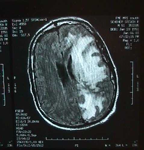
This was the initial presentation of ADEM on Jan.6th, 2003. The white area is
the affected portion of the left brain hemisphere. Persons with this much
involvement usually die. Note that the left brain hemisphere is on the right in this
image, and that it is putting pressure on the right hemisphere which is left in this
image.
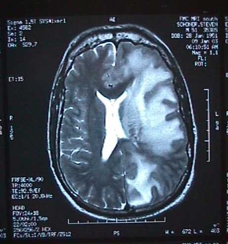
Jan. 9th, 2003. ADEM had advanced and I am in a coma. The corpus collosum
and right brain hemishpere are compressed by brain swelling. By this time ADEM
has progressed to the brain stem, and both sides of the occipital lobe
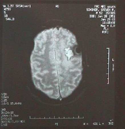
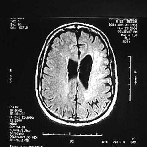
Jan. 29 th, 2003. 19 days after the brain biopsy. White area and black spot are
the results. Three fragments, nearly a cubic inch, of the left hemisphere were removed.
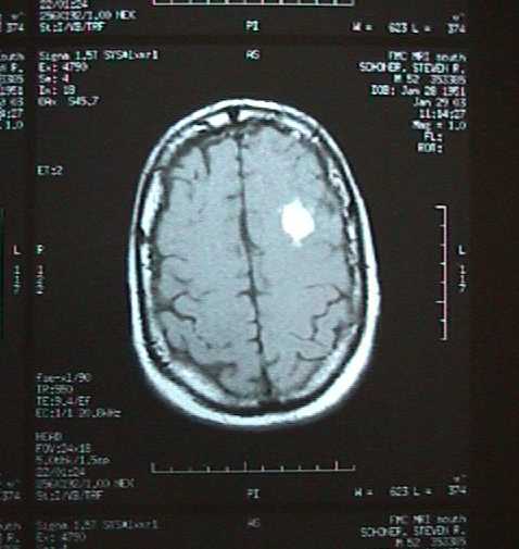
Jan. 29 th, 2003. Another view of the biopsy area. White area and black spots
are where the brain matter has been removed.
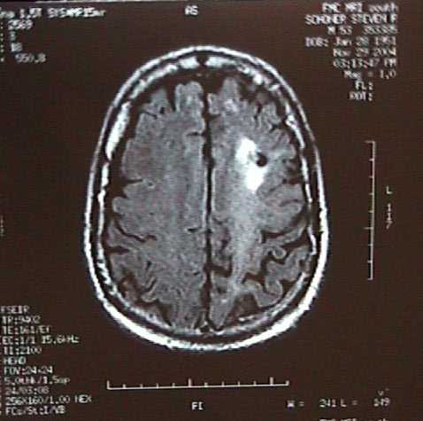
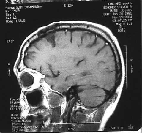
Nov 29th, 2004. Brain biopsy site is clearly visible in the white area of this image.
Nov. 29, 2004. Another image of biopsy Note the white area, and a
diffuse white haze around it, residual ADEM related demyelination.
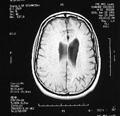
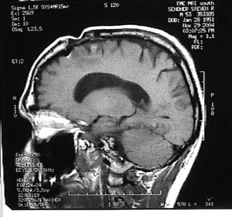
Nov. 29, 2004. Image showing brain shrinkage and abnormally enlarged.
corpus collosum on one side. Note dividing line between the two hemispheres.
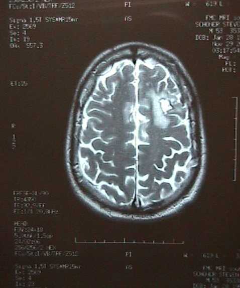
Nov. 29, 2004. Note the enlarged corpus collosum, and a diffuse white haze
around it-- Residual ADEM related demyelination.
Nov. 29, 2004. Another image of biopsy from left side. No brain in
dark wedge up and to the right of the eye socket where brain was
removed in biopsy. Skull above it is dark in this image where it was removed.
Nov. 29, 2004. Image of the dent in my head in cross section. White band
is my skin. Skull below was trepanned, and a 3" x 2.5" piece of skull removed
for the biopsy. Three samples were removed from this area.