Slides from my brain
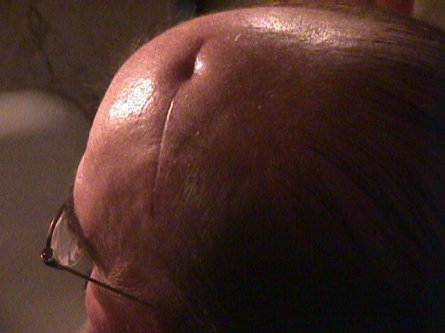
Photo 04/02/03
Having suffered life threatening acute disseminating encephalomyelitis (ADEM)-- Steve Schoner's brain biopsy scar shortly after his one month stay in the hospital.
MRI SCANS 01/06/03-11/29/04 showing extent of ADEM involvement=> MRI of brain
The purpose of this page is to present not only these remarkable biopsy slides taken from my brain, but to provide information links about this very rare disorder that is related to other demyelinating conditions, such as MS, Transverse Myelitis, as well as a diverse range of abnormal demyelination conditions.
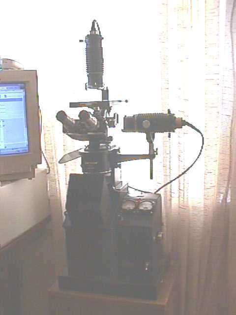
My Unitron Metallographic BU-11 microscope used to take the images of slides. This microscope can be used to examine metals such as in meteorites, and for petrologic studies for which I use it most. However, I have found that it also works very admirably, even with my limited experience with histology slides as with the following slides clearly show.
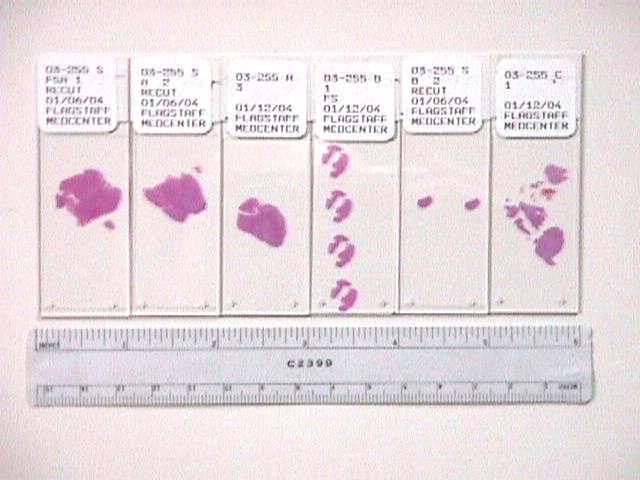
Six thin section histologic slides provided to me by the Pathology Department of the Flagstaff Medical Center after a substantial brain biopsy from the left frontal lobe of my brain done on January 10th, 2003 while I was deep in a coma. The area removed was described as Part A) 1.5 cm x 1.5 cm x 2.2cm; Part B) Fragments .3 - 1.0 cm; Part C) Several grey and yellow tissue pieces 0.6 cm x 1.5 cm x 1.7 cm and 0.4 cm x 0.8 cm x 1.3 cm. Slides as shown were made from the biopsy sample and the bulk of this sample remains with Flagstaff Medical Center. The official medical report indicates that the demyelinating condition was attributed to ADEM. But even then this diagnosis is uncertain as typical ADEM affects both hemispheres of the brain rather than one hemisphere, such as was in my case. What is certain is that an acute demyelinating condition was well under way on the left hemisphere of my brain when I was entered the hospital on Jan. 6, 2003
The following images were taken with my personal equipment, and the interpretations of what is seen in these slides are not contingent or relating to the official Pathology report, those of FMC, and or the neurosurgeons involved. And at this point in my recovery, I do not make any claims that my observatons are medically accurate, other than to say that they appear to relate to the information I gleaned in the resources listed at the end of this page. I will, however update this page once I can review the 76 page official Anatomic Pathology Report that pertains to these slides, and what they reveal.
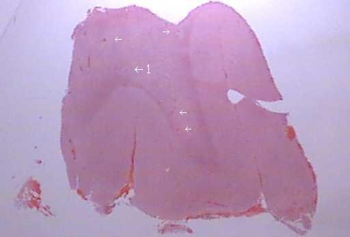
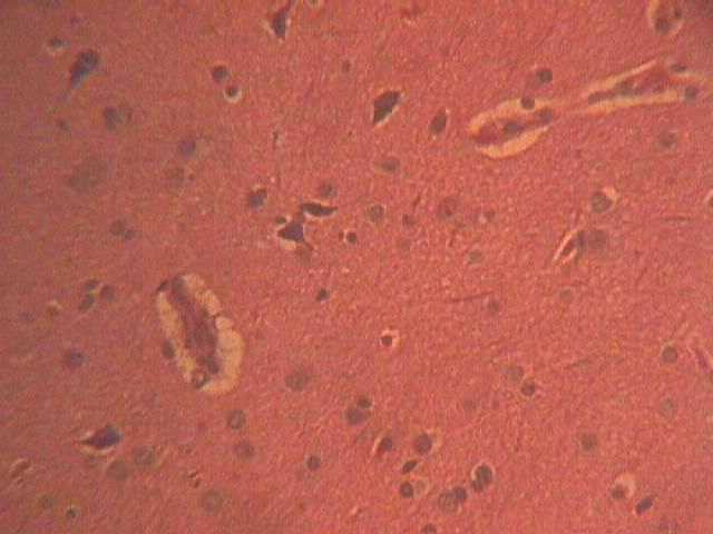
Brain section (A-2) showing white and gray matter. The largest dimension of this section is 3/4" (2 cm). There are several areas surrounding minor hemorrhages, showing as dark areas that are shaded lighter than the surrounding tissue. Arrows point to areas where demyelination has occurred.
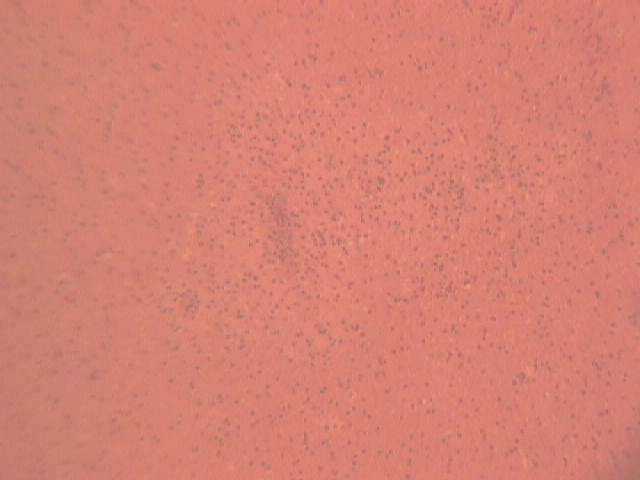
Close up at 50X of one of these lighter areas in the previous slide, showing dark area of hemorrhage surrounded and very much confined to the area of glial cells in area of demyelination from viral infection. 50X
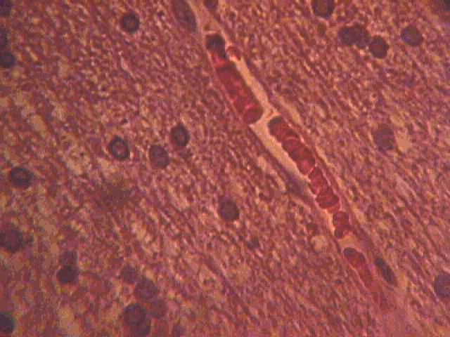
Enlargement of very small capillary with blood cells stacked like coins. Area surrounding it is gray matter with glial cells, astorcytes and oligodendrocytes. 400X
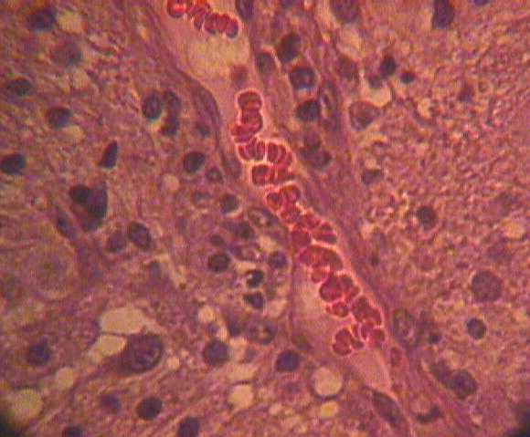
Enlargement of very small capillary with blood cells nearly one by one. Area surrounding it is white matter with glial cells, astorcytes, oligodendrocytes, macrophages and monocytes. Macriogages and monocytes are the white masses outside of the vein and are causing demyelination of the white matter. 400X
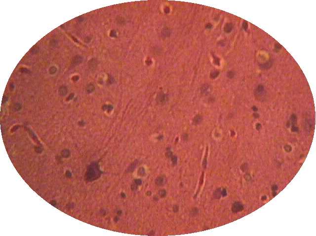
Cells, damaged in the cut edge of brain sample showing disturbed axions, glial cells, oligodenrocytes, and red blood corpuscles. 400X
Virchow-Robin space surrounding blood vessels within demyelinated, diffusely edematous white matter. 400X
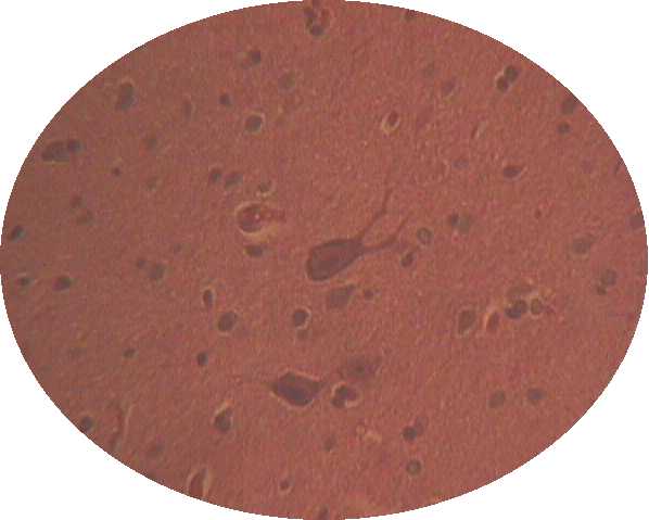
A very nice 500X photo of a branching Purkinje cell and neuron below it in affected white matter.
Another image of neurons with axions, Purkinje cells, and extremely small capillaries. 500 X
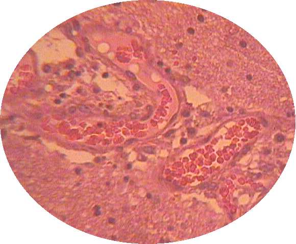
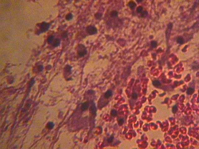
This incredible 300X image is of a spiral blood vessel with red blood cells within. The white areas are macrophage cells, and these have permiated the vessel walls penetrating white matter causing demyelination. The actual size of this view is > .1 mm.

Photo, from front page, AZ Daily Sun, July, 26th, 2003
I would like to extend my appreciation to members of the Flagstaff Medical Center Pathology Department for preparing and presenting to me these re-cuts from original tissue extracted from my brain on January 10th of 2003. The biopsy report described this as a "large brain biopsy" done when I was in a coma from this debilitating and nearly fatal condition. They did not expect me to survive. But, thank God I did, and am on the road to recovery. Needless to say, there are however certain disabilities that remain, and in light of this my former vocation as a computer technician is in question. But these slides have sparked my mind to study this rare condition, at least in thought and personal interest. That I survived this condition is a miracle, and in my current state of mind, looking at my own brain, to see and comprehend the things that I am seeing is awesome and inspiring-- Even more I think than I would feel if in space, or standing on the moon. Someday, perhaps I will write in layman's terms a story detailing this experience and what is currently known about this strange and rare disease. But for now I invite you to search the links that I have found.
To contact me, my e-mail is: steve_schoner@yahoo.com
ADEM INFORMATION LINKS:
Demyelinating Diseases-- extensive links to resources regarding this condition. Covers ADEM, MS, and other demylenating conditions. => Demyelinating Diseases
Internet Handbook of Neurology-- A very informative site that will provide excellent resources on the many conditions that can affect the brain. => Internet Handbook of Neurology/
Demyelinating Disease => Demyelinating Disease
The Myelin Project => The Myelin Project
ADEM at Contact a Family => Contact
Neurology India, ADEM-- An excellent abstract with resources on ADEM in children and adults. => ADEM
Neuropathies With Abnormal Myelination => Abnormal Myelination
Demyelinating Diseases of the Brain => Demyelinating Diseases
Demyelinating Disease (immune mediated or viral caused) => Demyelinating Diseases
Transverse Myelitis Association ADEM Forum-- A online forum providing personal experiences and resources on ADEM and other immune mediated diseases of myelination. => ADEM Survivors Forum
Fulminant Acute Disseminated Encephalomyelitis -- ADEM research in Turkey-- A very interesting abstract.=> ADEM
Because there seems to be a relationship between some cases of ADEM and multiple sclerosis with accompanying seizures, I have included the following links:
MS AND SEIZURE LINKS:
National Multiple Sclerosis Society information on seizures=> NMSS
eCureMe information on seizures=> eCureMe